Endoscopic Spine Surgery
Direct visualization and treatment of nerve compression through a sub-centimeter incision, with same-day discharge.
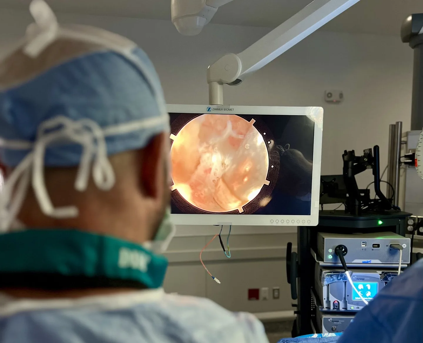
The Least Invasive Way to Treat a Pinched Nerve.
Endoscopic spine surgery uses a high-definition camera and specialized instruments delivered through an incision smaller than your fingertip, reaching the source of nerve compression while leaving the surrounding muscle and bone largely undisturbed.
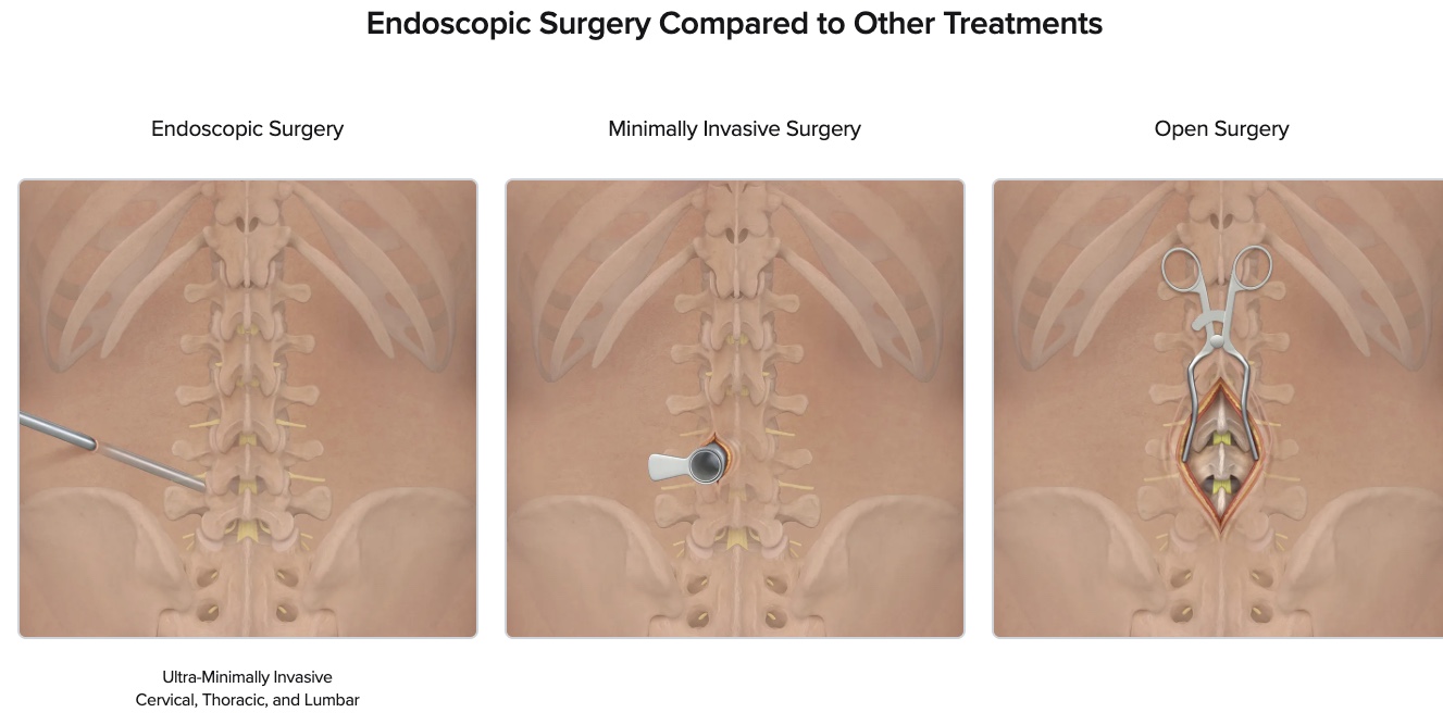
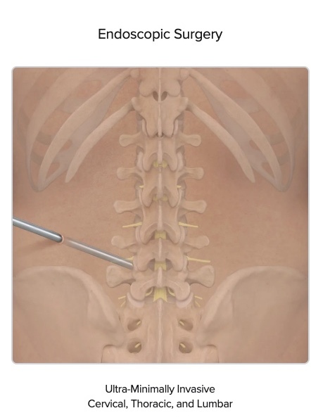
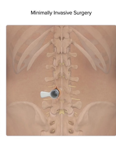
Advantages of the Endoscopic Approach
Sub-Centimeter Incision
A small working channel reaches the disc and nerve directly, with no muscle stripping.
Outpatient Procedure
Most go home the same day and resume light activity within days.
Targeted Relief
Direct visualization of the compressed nerve allows precise removal without disturbing nearby anatomy.
Preserved Motion
Fusion is avoided in appropriate patients, so the spine keeps its natural movement.
Foraminal Stenosis and Avoiding Fusion
Nerves leave the spine through a tunnel on each side called the foramen. When a nerve is compressed inside that tunnel, either by a disc fragment or by the narrowing that comes with arthritis, the problem is called foraminal stenosis or a foraminal disc herniation, and it has long been one of the harder problems in spine surgery to solve without a fusion.
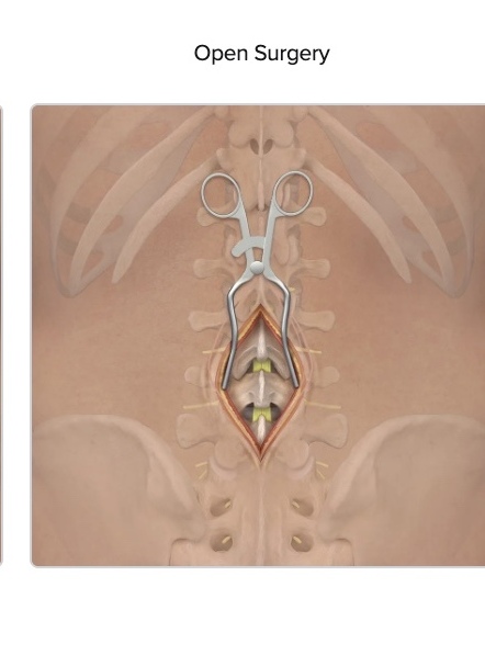
The reason is anatomy. The facet joint, one of the paired joints at the back of the spine, forms the back wall of the foramen. Without an endoscope, seeing the nerve where it is compressed has usually meant removing that joint in order to reach it, and removing a facet joint destabilizes the segment, which then has to be fused. For many patients with compression in this location, fusion was the operation on offer. The exposure required it even in cases where the underlying problem did not.
An endoscope changes the angle of approach. The instrument enters on a trajectory that looks directly into the foramen, so the nerve can be seen and the pressure taken off it without removing the bone that holds the segment together. The segment stays stable, and the larger operation is often unnecessary.
What Endoscopic Surgery Can Treat
The endoscopic approach is a way to reach the spine, applied across several problems where a nerve is compressed and the pressure can be relieved with little disruption to the surrounding tissue.
Herniated Disc
A disc fragment pressing on a nerve root is removed through a dime-sized incision, relieving the leg pain it causes.
Spinal Stenosis
Narrowing that crowds the nerves can be opened in appropriate cases, taking pressure off the nerves while preserving the surrounding bone and muscle.
Nerve Root Compression
The bony opening a nerve root passes through can be widened to free a pinched nerve, an endoscopic foraminotomy.
Dr. Hirsch is board-certified and Yale-trained. Read his full background.